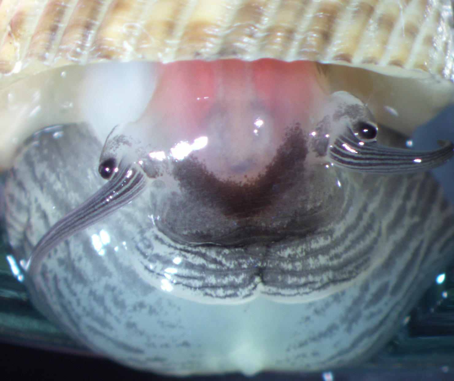
Figure 1: Soft body of Nerita undata. Image by Rebecca Loh.
Introduction
Table of Contents
The undate nerite (also known as waved nerite) [1] is a species of marine snail [2,3] that has a wide distribution across tropical and temperate zones [4]. Nerita undata used to be considered a single species, but it has come to light that what we consider as one species may in fact be multiple species [5]. Hence, neotype has recently been designated to stabalise its taxonomic status [6, 7]. Externally, this species may looks unremarkable, but many do not realise that its charisma "comes from within"; its soft body. Most remarkably, it is a member of the only snail family whereby the females of many species have a special storage organ - the crystal sac [8]! Female undate nerites store lime crystals in this storage organ, and the crystals are used to strengthen their egg capsules.
Reproduction
Sexual dimorphism
Did you know that the penis of nerites is located on their head? [9] Sexes within the undate nerites are separate and there is no obvious dimorphism in terms of shell characters. However, the male head organ (penis) is located between the eyes and tentacles!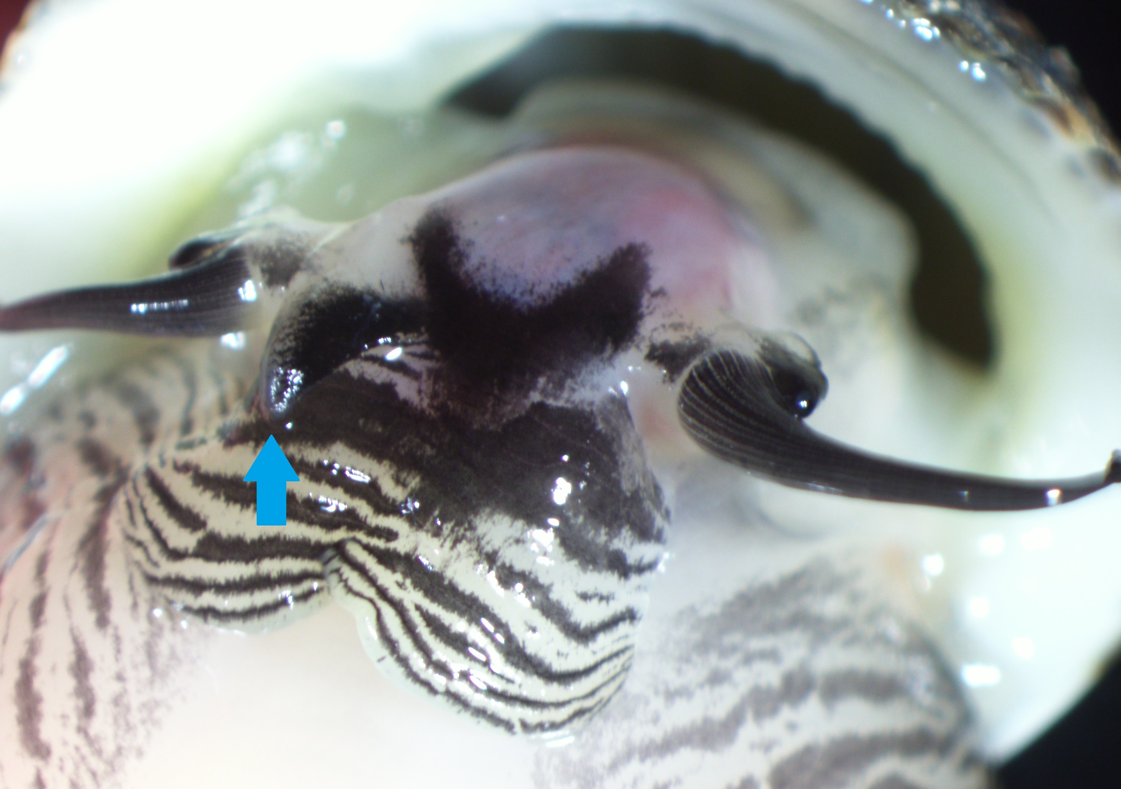 |
This dimorphism can aid sex determination in the field without hurting the animal, but this requires some patience because nerites are generally shy and will retract into its shell if you pry their shells open too suddenly!
Mating behaviour
The mating process of a close relative of undate nerites, Nerita funiculata Menke, 1851, has been observed and recorded in a study (read about the juicy account in this paper) [9]. But the process is rather different for undate nerites (personal observation).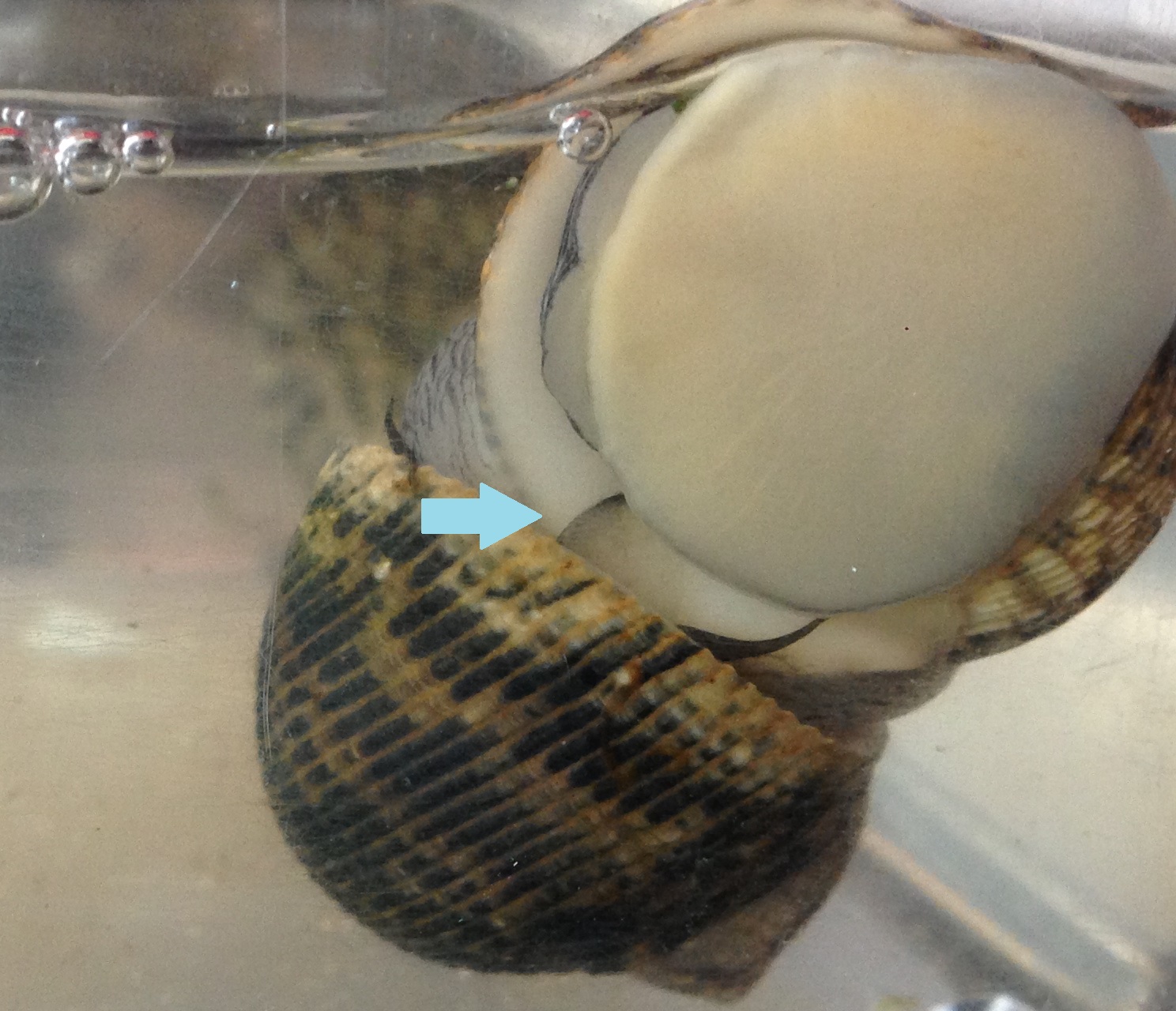 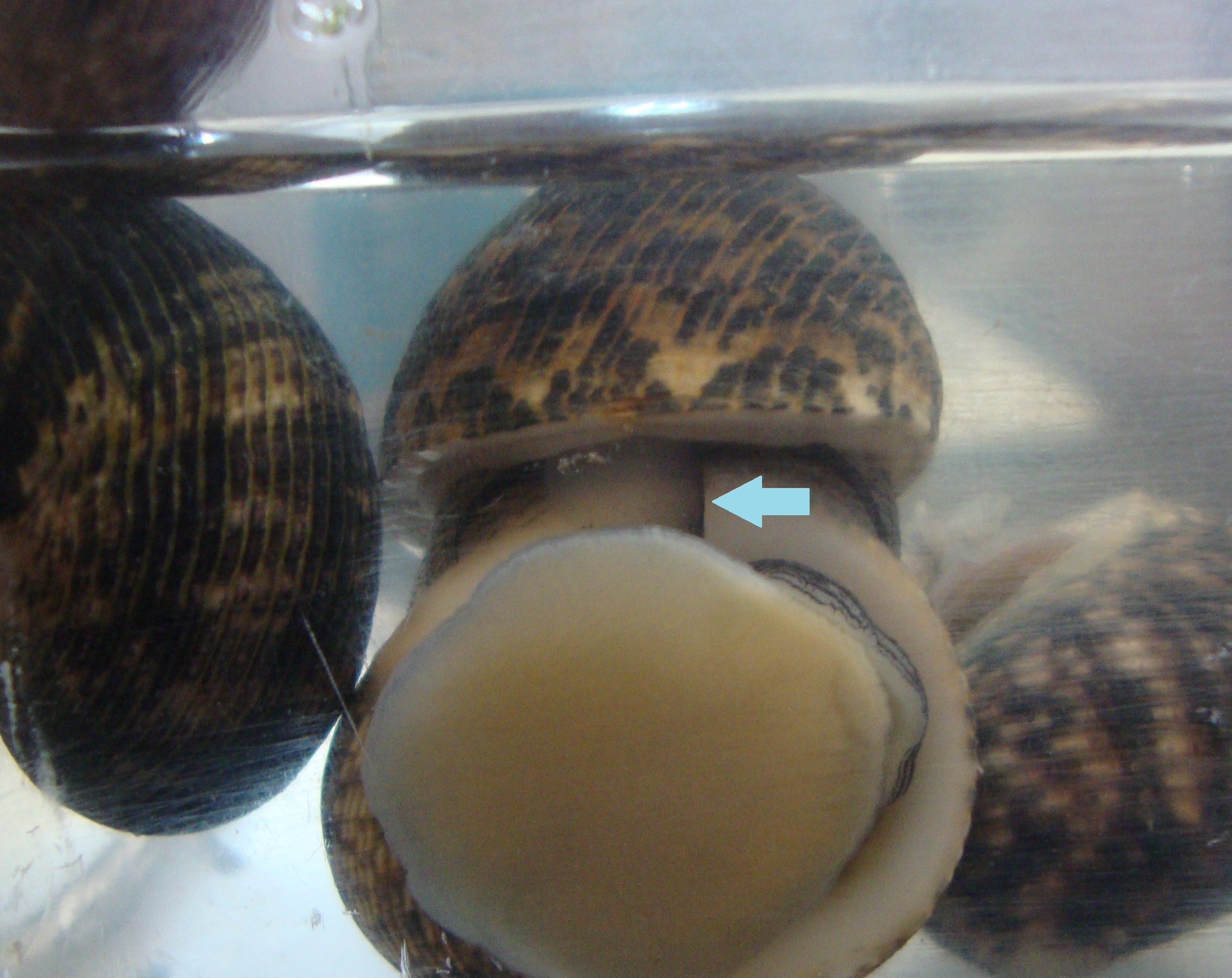 Figure 3: Photographs of Nerita undata pair copulating. Arrows: penis, which extends into the shell of females. Image by Rebecca Loh. |
Before mating, the undate nerite male climbs onto the shell of females and uses the penis to make contact with the right side of the female soft body (personal observation). A cycle follows: penis extension, making contact with female, retraction and extension again (shown in Videos 1 and 2). Eventually, the male genitals reach into the right side of the female shell and stays there for an extended period of time, during which white masses (probably the sperm-containing spermatophores, Figure 4) can be observed sliding through translucent the male organ towards the female (shown in Video 2). After copulation, the male retracts its penis and crawls away.
Video 1: Mating behaviour of Nerita undata prior to copulation. Video by Rebecca Loh. |
Video 2: Mating behaviour of Nerita undata, including courtship and copulation. At 3:11, white masses (spermatophores) were observed sliding from male to female. Video taken and annotated by Rebecca Loh |
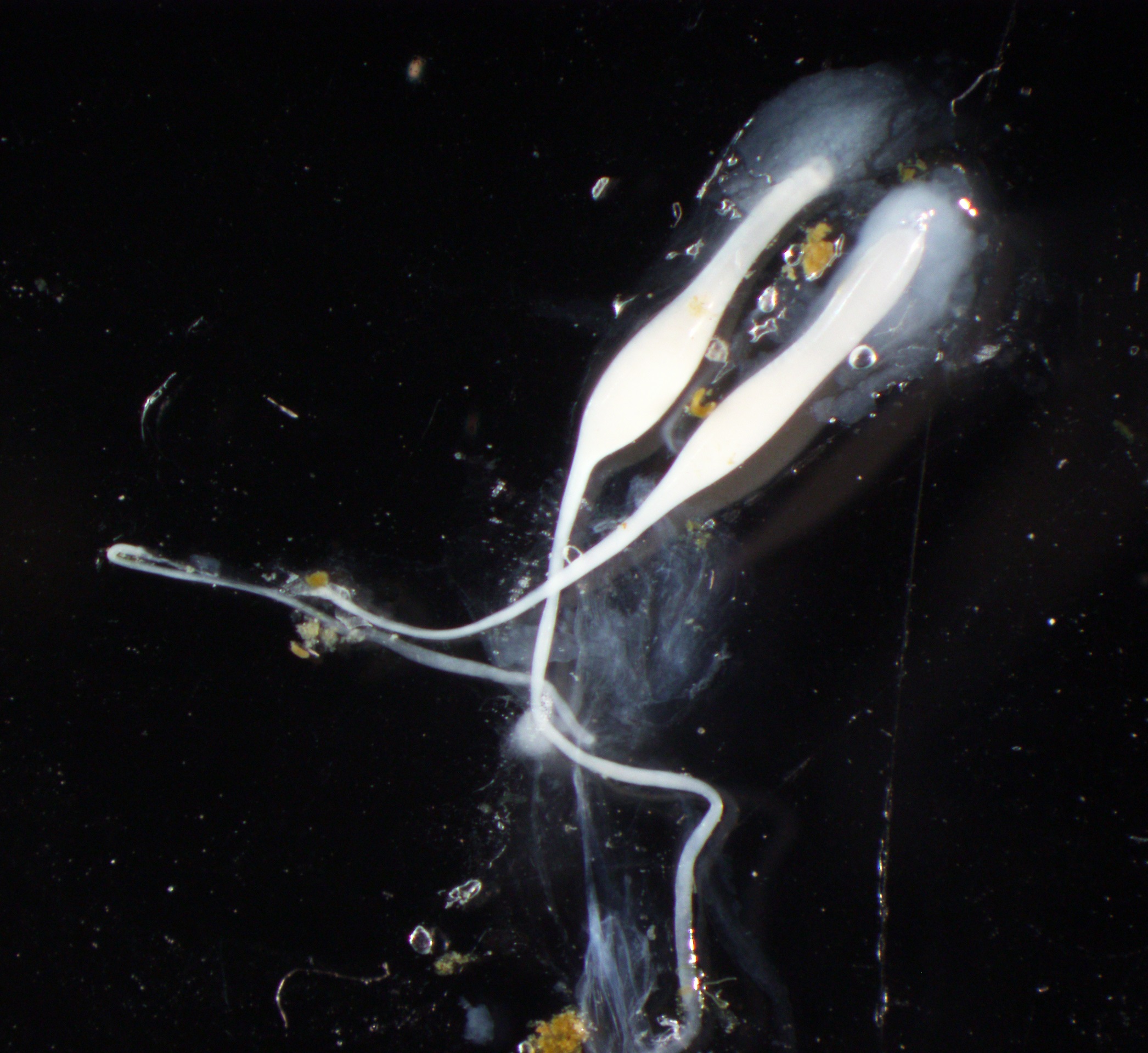 Figure 4: Photograph of spermatophores of Nerita undata. Image by Rebecca Loh. |
Reproductive anatomy
What is going on inside the snail during copulation and reproduction? As mentioned, the male genital is located on the head of nerites (Figure 5: P, blue box). During copulation, spermatophores discharged from the prostate gland (Figure 5: pg) travels to the penis via the cilliated groove (Figure 5: cgr) [9]. Figure 5: Illustration of the male genital ducts of Nerita funiculata Menke 1851, closely-related to Nerita undata. Legend: aux, auxiliary glands; cgr, ciliated groove; p, penis (blue box); pg, prostate gland; sv, seminal vesicle; t, testis; te, tentacle; vd, vas deferens. Access the full paper here [9]. Image extracted and adapted by Rebecca Loh, used under CC BY-NC-SA 4.0. Image from the Biodiversity Heritage Library. Digitized by BioStor. | www.biodiversitylibrary.org |
Female nerites receive spermatophores and store them in the spermatophore sac (Figure 6: sps) [9]. Prior to fertilisation, sperms are released from the spermatophores and they travel to the oviduct (Figure 6: ov) where fertilisation takes place [10]. Larvae are encapsulated in egg capsules, which travel through and exit the capsule gland (Figure 6: cg), and becomes attached to hard substratum [10].
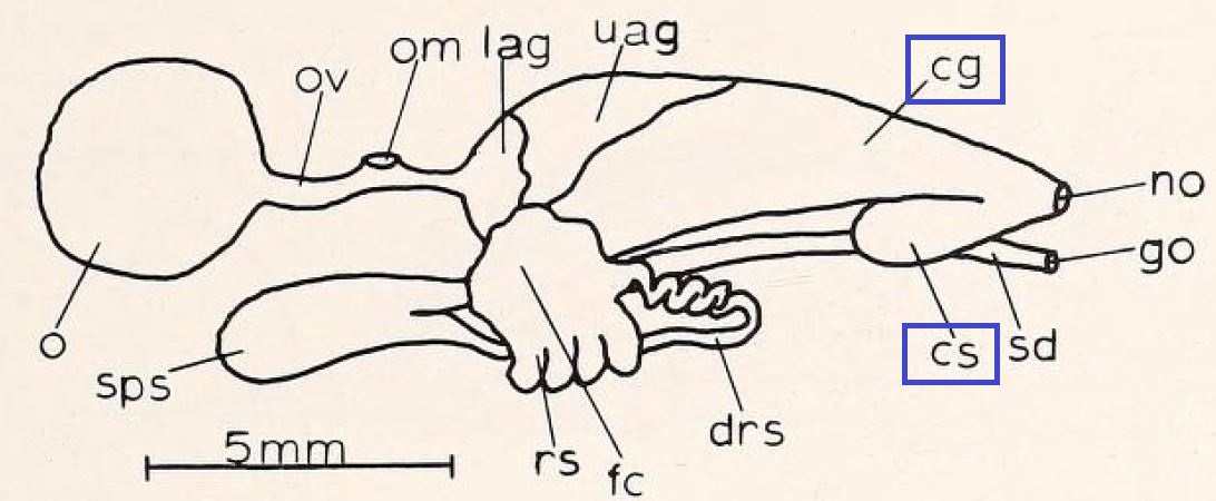 Figure 6: Illustration of the female genital ducts of Nerita funiculata Menke 1851, closely-related to Nerita undata. Legend: cg, capsule gland or ootype (blue box); cs, crystal sac or reinforcement sac (blue box), drs, duct to receptaculum seminis; de, ductus enigmaticus; fc, fertilisation chamber; go, genital opening; lag, lower albumin gland; no, nidamental opening; o, ovary; ov, oviduct; om, opening of oviduct into mantle cavity; rs, receptaculum seminis; sd, sperm duct; sps, spermatophore sac; uag, upper albumin gland. Access the full paper here [9]. Image extracted and adapted by Rebecca Loh, used under CC BY-NC-SA 4.0. Image from the Biodiversity Heritage Library. Digitized by BioStor. | www.biodiversitylibrary.org |
Most remarkably, nerites (Family Neritidae) are the only family of snails whereby the females of some species have the crystal sac/reinforcement sac (Figure 6: cs) which stores minerals that are used to harden the surface of egg capsules [10]. As the capsules exit the capsule gland, minerals are sprinkled all over its surface [9]. Read on to find out more about egg capsules!
Egg capsules and reinforcement materials
Nerites encapsulate their larvae in membranous egg capsules, which they lay on hard substratum along intertidal areas [11]. Egg capsules of undate nerites ranges from 2.8-3.2 mm in diameter and can contain not just one, but between 15-37 embryos [11]! When the time is right, it was observed in another nerite species (Dostia violacea) that the capsule cap would swing open under pressure and release larvae into the water column [11].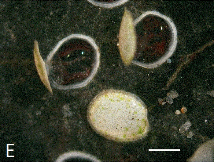 Figure 7: Photograph showing two opened egg capsules and one intact capsule of Dostia violacea. Scale bar = 1mm. Image from Tan and Lee (2009) [11] with permission. |
Certain nerite species use minerals like sand or lime (also known as calcium carbonate) crystals to reinforce the surface of their egg capsules, supposedly to prevent desiccation, deter predation, yet enabling gaseous exchange at the same time [11]. It has been suggested that the egg capsule surface morphology is species-specific and could be useful in taxonomy, especially for distinguishing closely related species [10, 11]. This is supported by a study [11] on the egg capsules of six neritid species, which revealed that each has unique egg capsule morphology.
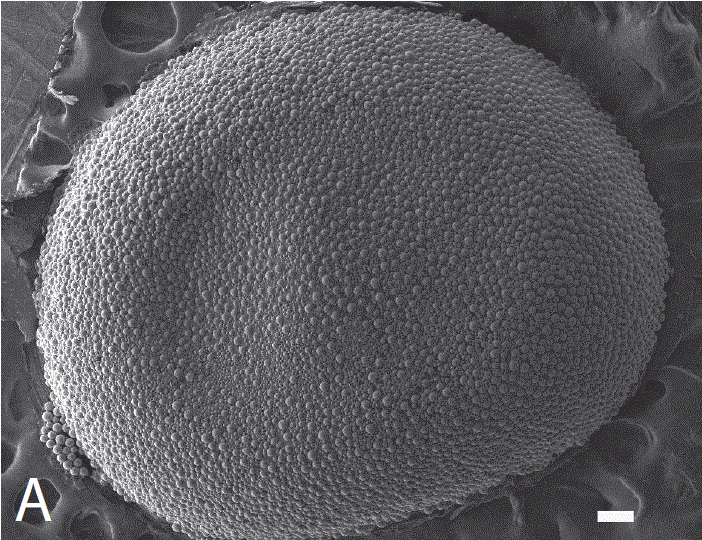
Undate nerites use calcium crystals to reinforce the surface of their egg capsules. Its crystals are spherical, and range from 20-60µm in diameter [11]. These calcium carbonate spherulites are loosely scattered in the centre of the cap and densely packed near the edges [11]. There is no observed patterns in the size distribution of spherulites.
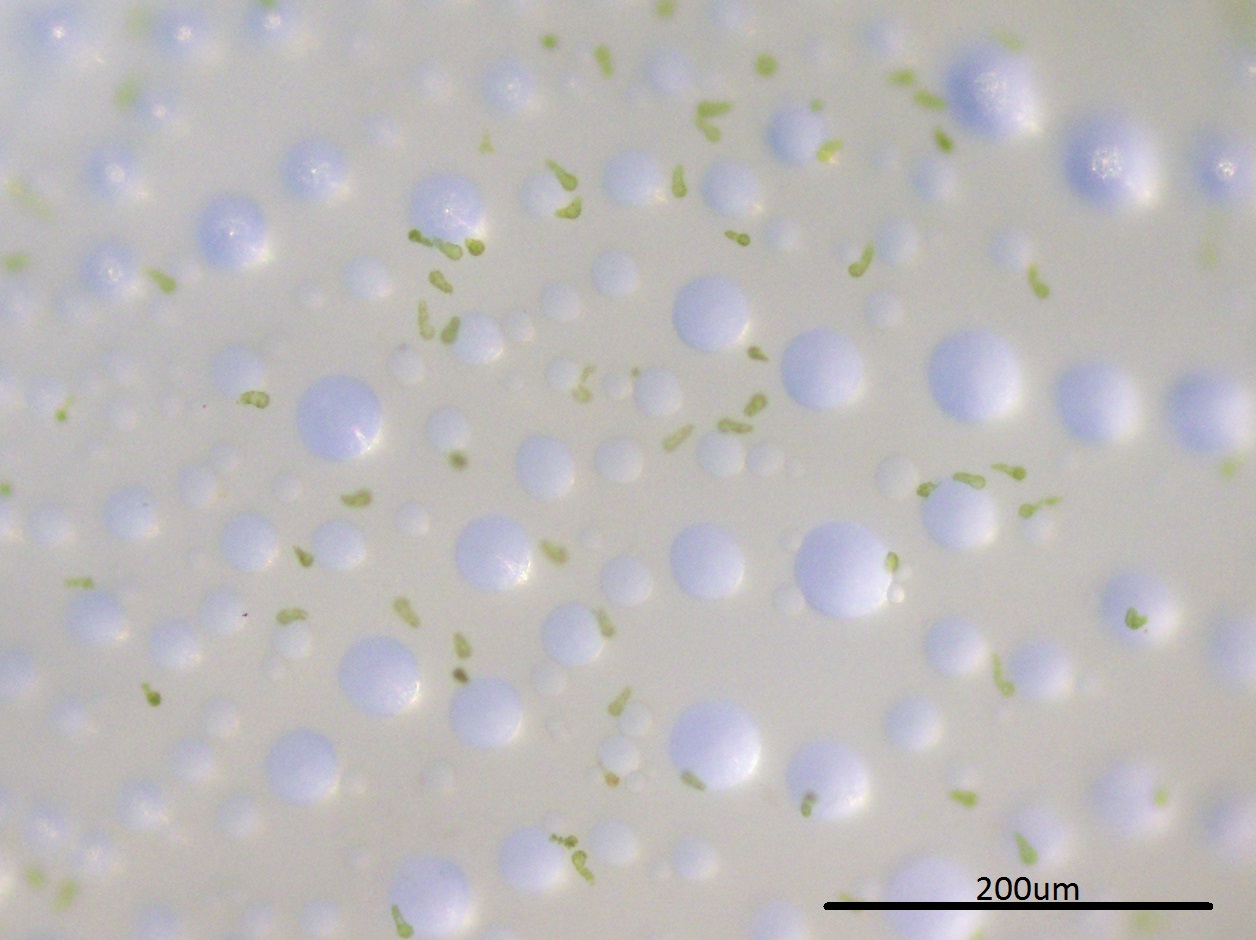 Figure 8: Photograph showing calcium carbonante spherulites on the egg capsule surface of Nerita undata, under the light microscope. Image by Rebecca Loh. |
Figure 9: Image showing calcium carbonante spherulites on the egg capsule surface of Nerita undata, when viewed under the Scanning Electron Microscope. Scale bar: 100µm. Image from Tan and Lee (2009) [11] with permission. |
Crystal sac - a mystery!
Calcium carbonate crystals or spherulites have been observed in the reinforcement sac (also known as the crystal sac) of the females. It is speculated that the crystals are stored in the crystal sac, and deposited on the egg capsules before they are laid [8, 12].You might be wondering: so how did the lime spherulites end up in the reinforcement sac? Well, the location that spherulites are produced and how they ended up in the reinforcement sac remains a mystery. There have been two speculations: The first is that spherulites were produced in the liver and transported through the alimentary canal before ending up in the crystal sac [8, 10, 12]. This was suggested by Andrews who claimed to have found spherulites in the liver (although no one confirmed this observation subsequently). The second speculation is that spherulites are formed by modifying lime particles (taken in with food) as they pass through the alimentary canal [9].
Either way, both speculations converge in that calcium spherulites that were formed were eventually transferred from the alimentary canal opening to the capsule gland opening and into the crystal sac. This is illustrated in Figure 10, blue arrow shows the path of calcium spherulites being transferred from the rectum (the end of the alimentary canal) to the crystal sac.
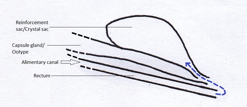 Figure 10: Illustratration of the path of calcium spherulites from the rectum (the exiting end of the alimentary canal) to the crystal sac. Image by Rebecca Loh. |
However, another study [11] rejected these speculations based on the observation that crystals in the crystal sac were too homogenous to be filtered out from rectal contents. They suggested that the spherulites were produced in another location which bypasses the rectum. But no alternative mechanism has been proposed.
Diet and Feeding
In general, nerites are herbivores [4] that feed on algae and diatoms. They feed by extending their snouts and their radula brushes and scraps the surface of substratum to gather food [13]. Undate nerites can be induced to feed by smearing algae (collected from rocks or seawalls) on substratum (personal observation).
Video 3: Nerita undata individual with its snout extended and its radula visibly brushing and scrapping the glass surface that has been smeared with algae. Video by Rebecca. |
In Mkomani, Monbasa, Kenya, it has been shown that Undate nerite populations exhibit feeding migration patterns that are size specific [14]. Migration starts at night (at around mid to ebb tide) and larger individuals would move down to their feeding levels while smaller individuals move up. individuals then return to their starting or resting positions with the flood tide, until the next nocturnal ebb tide comes [14].
Internal anatomy
Excellent observations and illustrations of the nerite anatomy have been documented by various researchers [9, 10, 14, 15], ranging from the digestive system to the alimentary canal. (Example of illustrations in Figure 11) However, the defining characteristics of nerites (Family Neritidae) remain to be the reproductive system and the presence of a crystal sac/reinforcement sac in the females of some nerites.
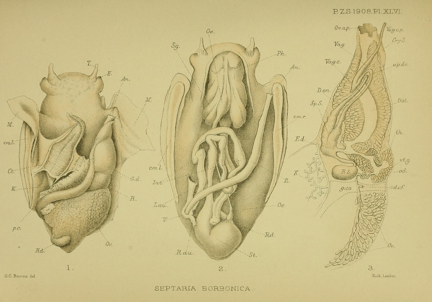 Figure 11: An example of detailed illustrations of the internal anatomy of Septaria borbonica, from a key anatomical study of nerites. From left to right: Respiratory system, alimentary canal, female reproductive system. Access the full paper here [15]. Image extracted and adapted by Rebecca Loh, used under CC BY-NC-SA 4.0. Image from the Biodiversity Heritage Library. Digitized by BioStor. | www.biodiversitylibrary.org |
Morphology
Species diagnostic characters
The diagnostic characters collated here have been adapted from various literature [4, 7, 16]. All photographs supplemented are that of two Nerita undata (live) specimens collected from St. John's Island, Singapore. The specimens follow closely to descriptions of the neotype recently designated [7].| General shape and colour |
|
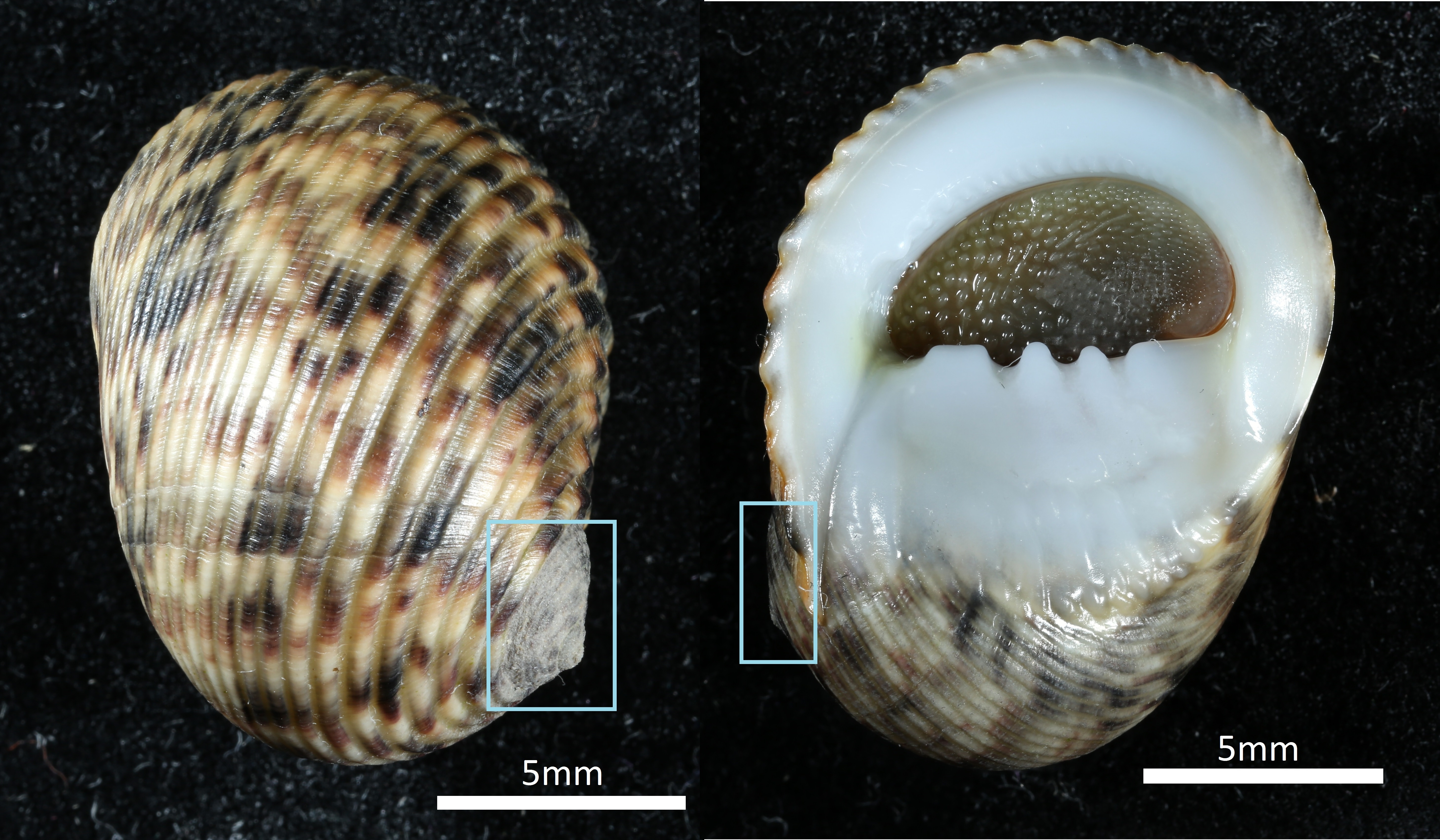 Figure 12: Dorsal view (right) and ventral view (left) of Nerita undata, shell width ~1cm. Boxed: spire. Image by Rebecca Loh. 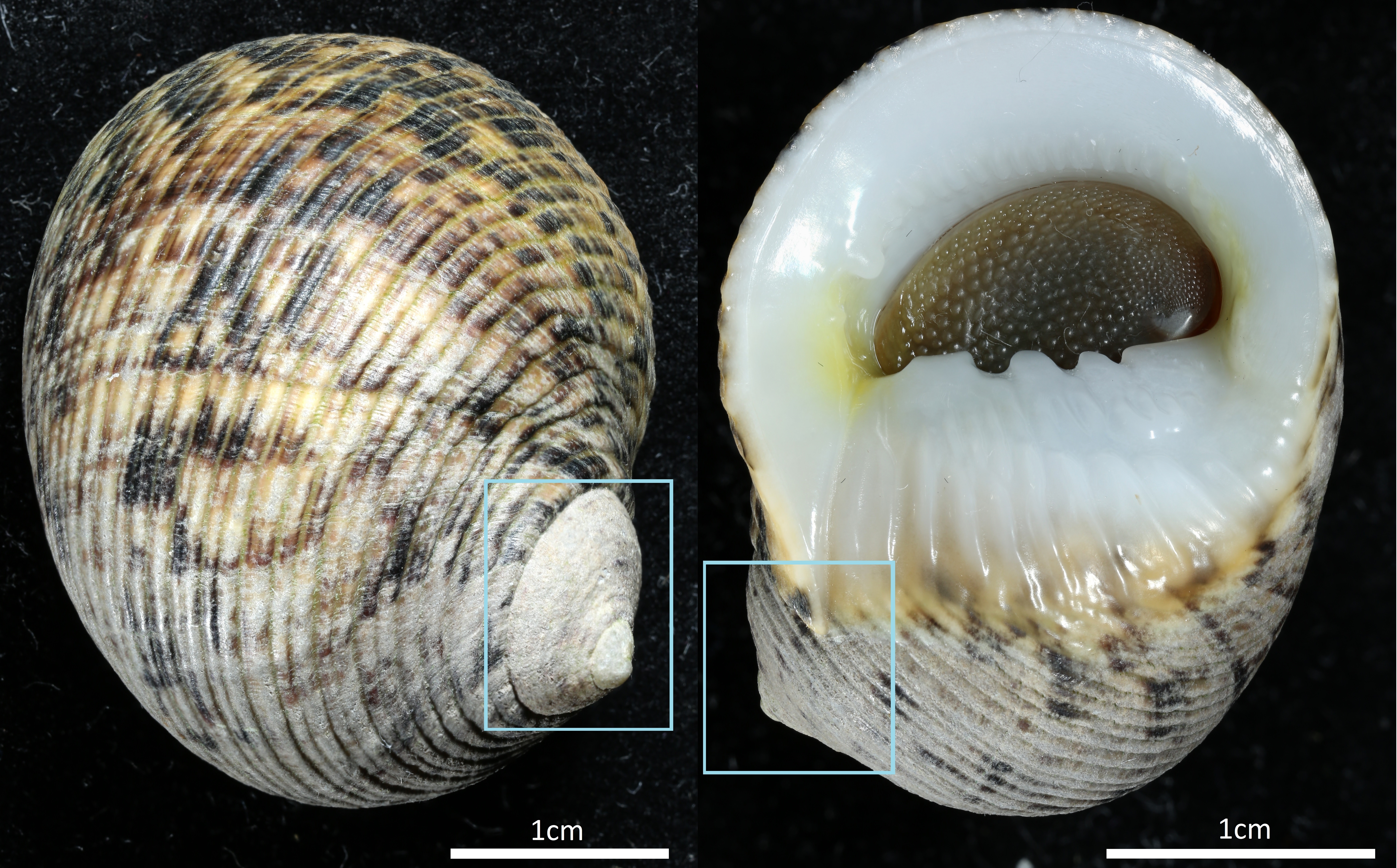 Figure 13: Dorsal view (right) and ventral view (left) of Nerita undata, shell width ~2cm. Boxed: spire. Image by Rebecca Loh. |
| Ribs |
|
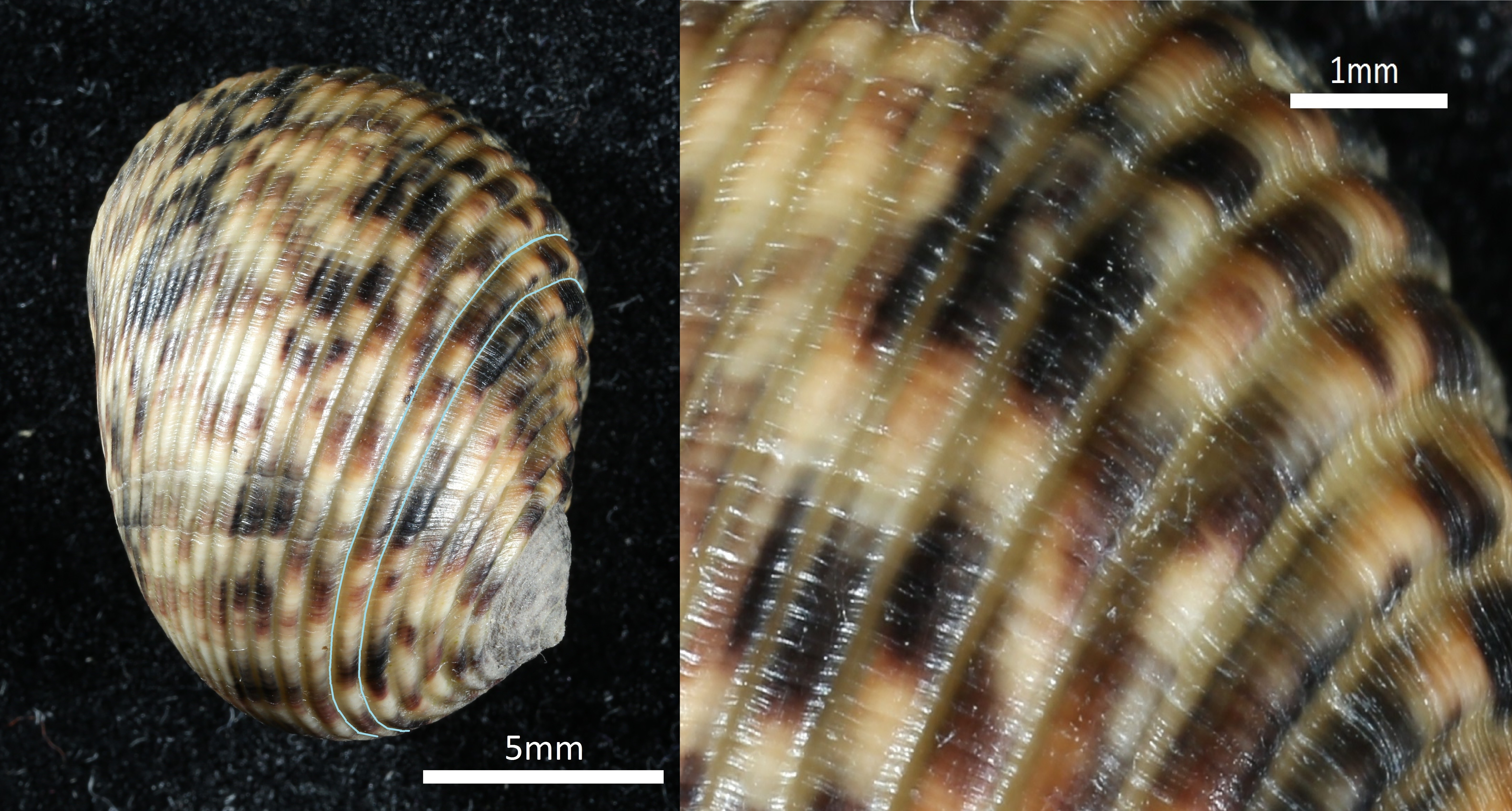
Figure 14: Dorsal view of Nerita undata, shell width ~1cm. Right: spiral rib outlined in blue. Left: enlarged view of spiral ribs. Image by Rebecca Loh. |
| Columella teeth |
|
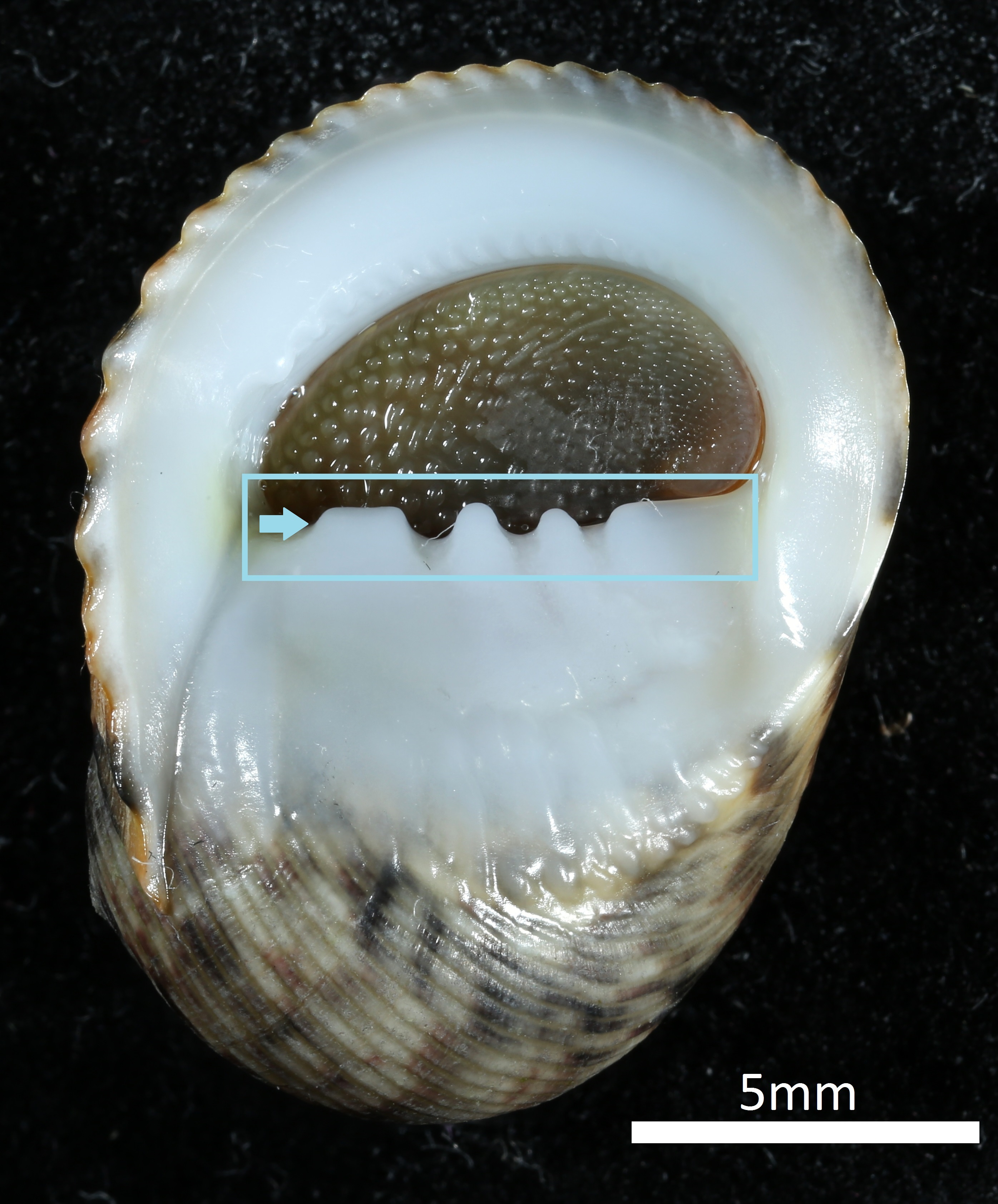 Figure 15: Ventral view of Nerita undata, shell width ~1cm. Boxed: columella teeth. Arrow: Large, squarish, upper columella tooth. Image by Rebecca Loh. |
| Parietal shield |
|
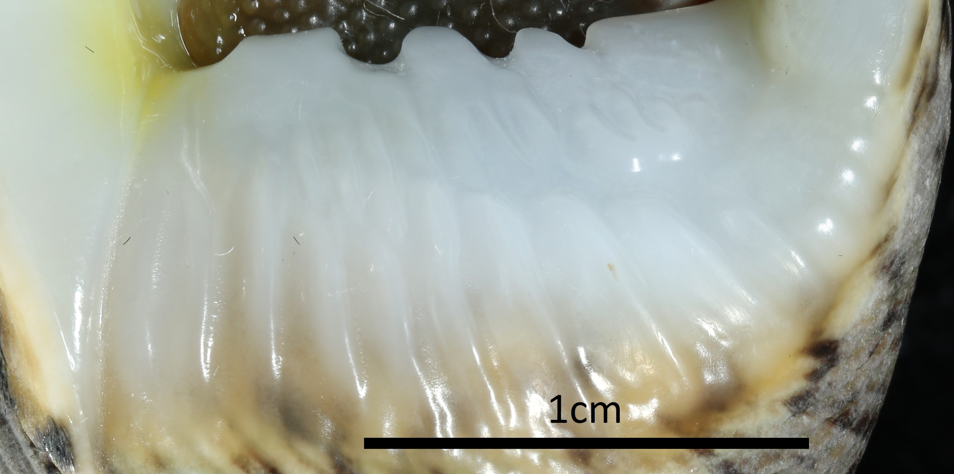 Figure 16: Ventral view of Nerita undata, shell width ~2cm. Boxed: wrinkled parietal shield. Image by Rebecca Loh. |
| Outer lip |
|
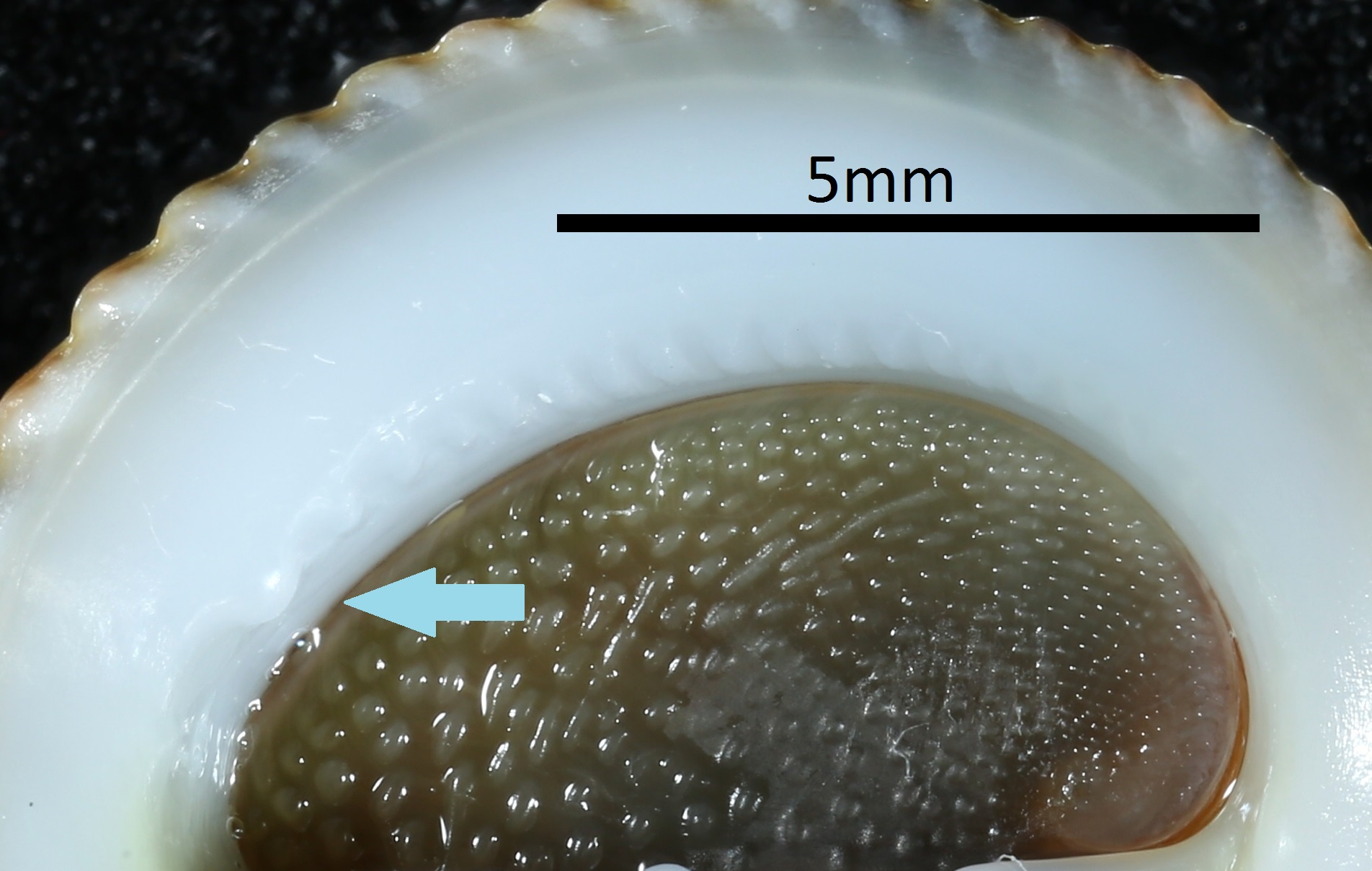 Figure 17: Ventral view of Nerita undata, shell width ~1cm. Arrow: prominent tooth on outer lip. Image by Rebecca Loh. 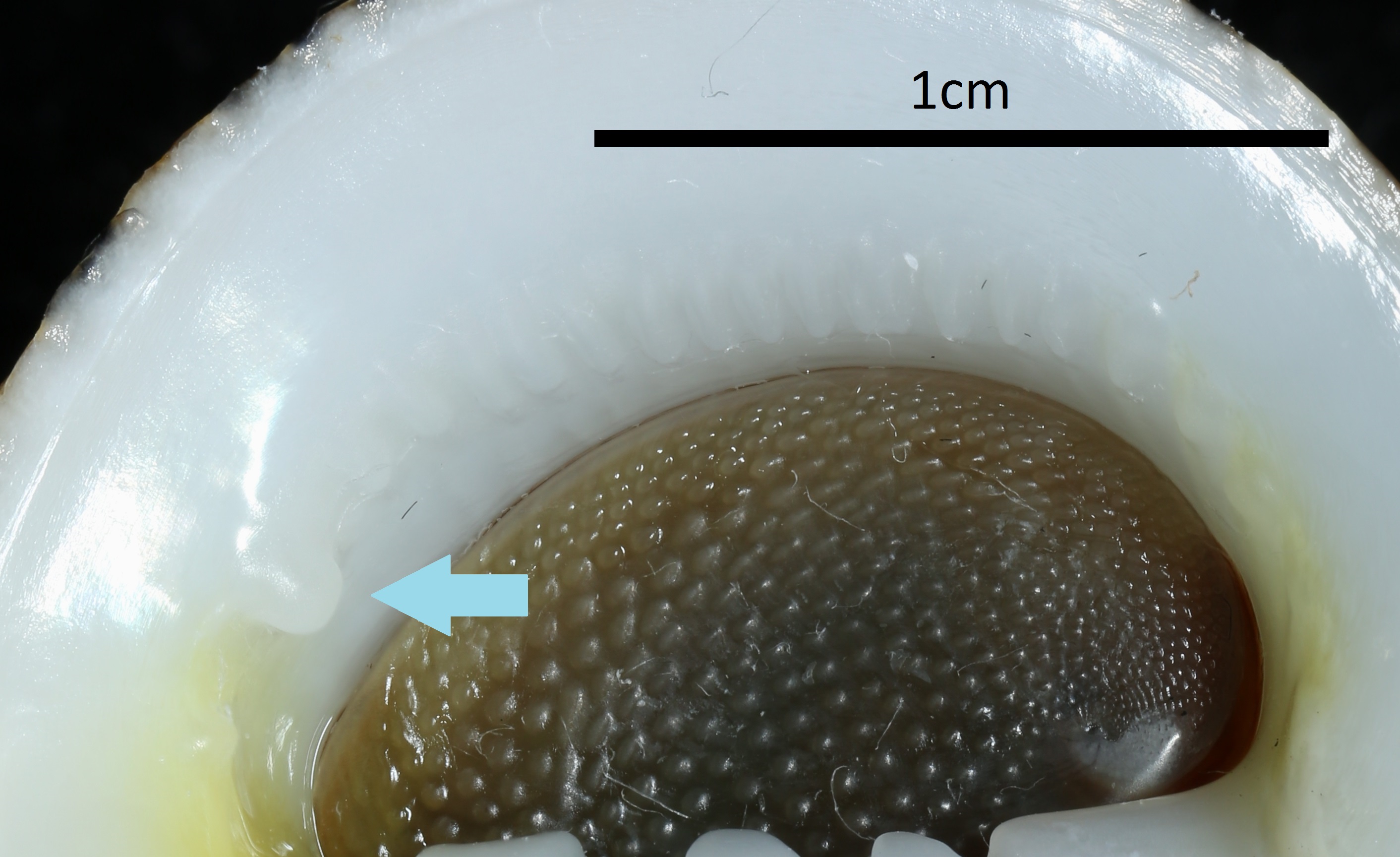 Figure 18: Ventral view of Nerita undata, shell width ~2cm. Arrow: prominent tooth on outer lip. Image by Rebecca Loh. |
| Aperture and operculum |
|
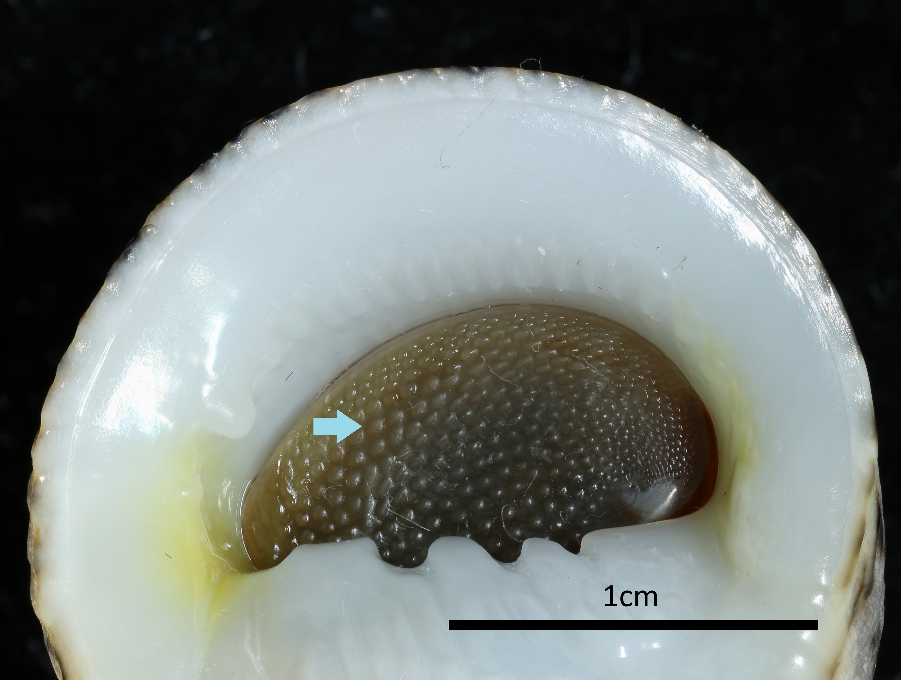 Figure 19: Ventral view of Nerita undata, shell width ~1cm. Arrow: pustule on operculum. Image by Rebecca Loh. |
| Soft body |
|
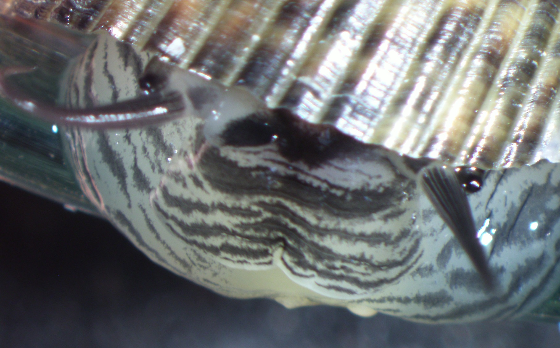 Figure 20: Soft body of Nerita undata. Image by Rebecca Loh |
Identification key
The following identification key was found on documents provided by the Food and Agriculture Organisation of the United Nations (FAO) [17]. However, it only allows differentiation between eleven neritid species, commonly found in the Western Central Pacific.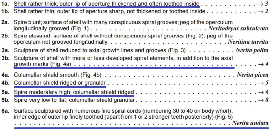 Figure 21: A section of the identification key to eleven nerite species of interest to fisheries of Western Central Pacific. Parts relevant to Nerita undata were underlined in blue. Species included in the original key: Nerita albicilla Linnaeus, 1758, Nerita chameleon Linnaeus, 1758, Nerita costata Gmelin, 1791, Nerita picea Récluz, 1841, Nerita planospira Anton, 1839, Nerita plicata Linnaeus, 1758, Nerita polita Linnaeus, 1758, Nerita squamulata Le Guillou, 1841, Nerita undata Linnaeus, 1758, Neritina turrita (Gmelin, 1791), Neritodryas subsulcata (Sowerby, 1836). Access the full version of the key from the FAO website here. Image extracted and adapted by Rebecca Loh, used according to website policy. Disclaimer: This is an adaptation of an original work by FAO. Views and opinions expressed in the adaptation are the sole responsibility of the author or authors of the adaptation and are not endorsed by FAO. |
Differentiate closely-related nerites
Have trouble differentiating closely-related nerite species? No need to fret. The following guide summarises the main differences between four nerite species that resemble undate nerites [4]. Key characters you can use to differentiate undate nerites from each species have been bolded.
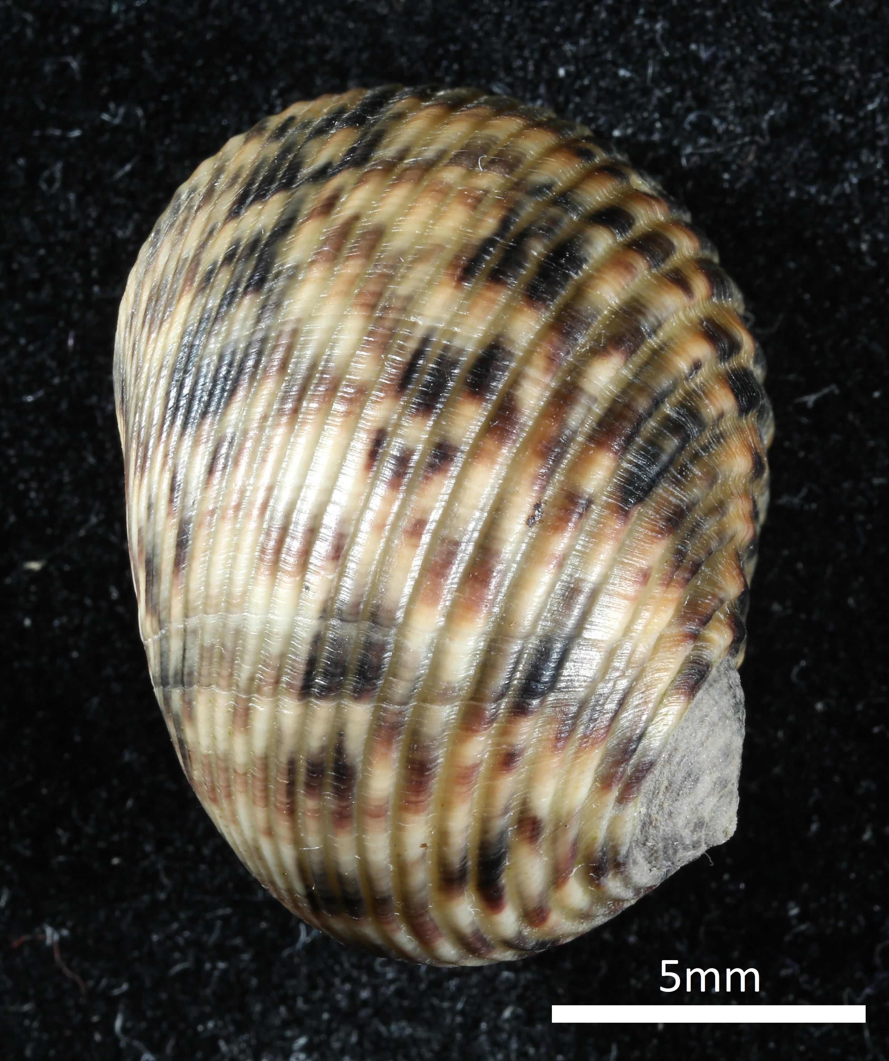 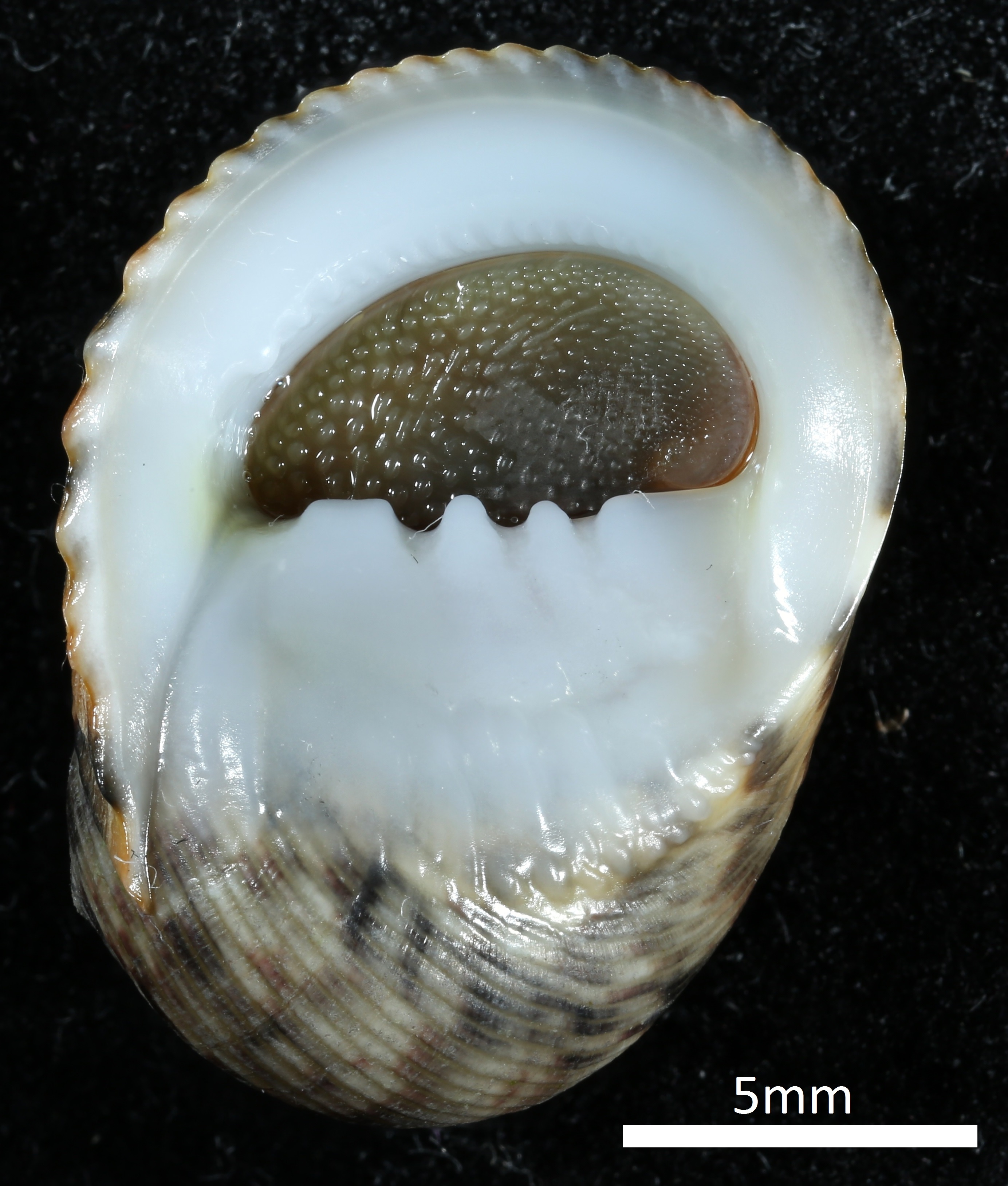 Figure 22: Dorsal (top) and ventral (bottom) views of Nerita undata. Image by Rebecca Loh |
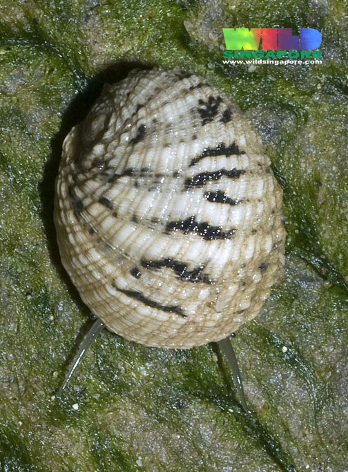 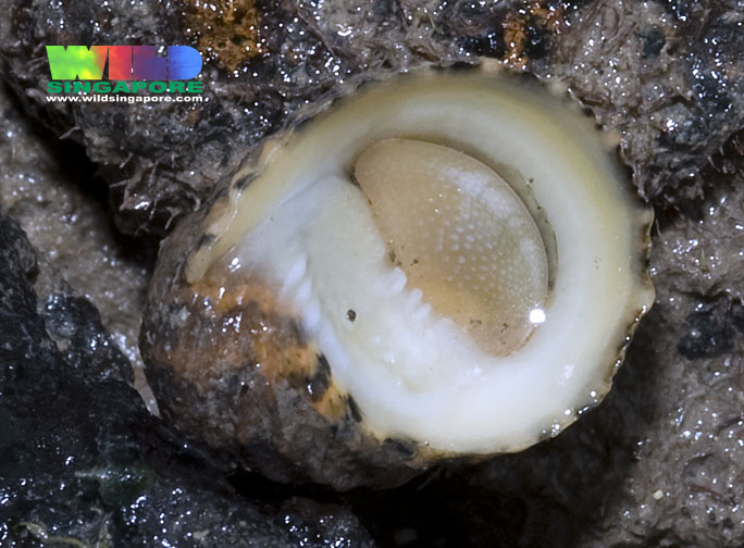 Figure 23: Dorsal (top) and ventral (bottom) views of Nerita histrio. Image by Ria Tan, used according to website policy. |
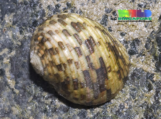 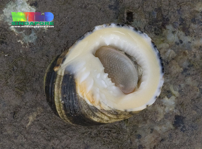 Figure 24: Dorsal (top) and ventral (bottom) views of Nerita chameleon. Image by Ria Tan, used according to website policy. |
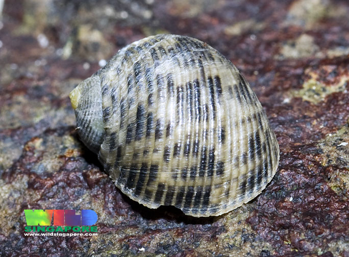 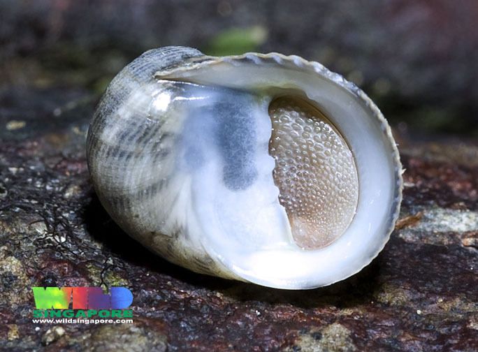 Figure 25: Dorsal (top) and ventral (bottom) views of Nerita grayana. Image by Ria Tan, used according to website policy. |
|
| Nerita undata Linnaeus, 1758 |
Nerita histrio Linnaeus, 1758 |
Nerita chameleon Linnaeus, 1758 |
Nerita grayana Recluz, 1844 |
|
| Shell sculpture (dorsal side) |
Low spiral ribs |
Rough, unevenly raised spiral ribs Axial sculpturing (perpendicular to ribs) prominent |
Raised spiral ribs Indistinct axial sculpturing |
Low spiral ribs Fine axial sculpturing |
| Spire |
Moderately high |
Low to nearly flat |
Low |
Moderately high |
| Columellar edge teeth |
3-5 teeth Uppermost tooth squarish |
Few, small obsolete teeth, that are centred |
2-4 small teeth, that are centred |
3-4 teeth Uppermost tooth squarish |
| Parietal shield |
Wrinkled White with varying degrees of yellow staining |
Smooth to small wrinkles White or pale yellow |
Several small pustules/granules, and wrinkled |
Smooth to strongly wrinkled Colour varies from white with yellowish tinge to yellow with orange tinge |
| Outer lip |
Dentate Prominent last tooth at upper end |
Dentate No distinct tooth |
Dentate 1 distinctly larger tooth at upper corner |
Dentate Prominent last tooth at upper end |
| Operculum |
Greyish Granules present Flat |
Flesh-coloured Granules present Somewhat convex |
Greyish Granules present Flat |
Greyish, usually with patchy beige coloration Small granules over entire surface Flat |
| Animal soft body |
Grey with black lines |
Sand-coloured with black lines |
Light grayish with black lines |
Creamy-white |
Shell mineralogy
As the soft bodies of snails grow, their mantle secretes minerals to enlarge their correspondingly [18, 19]. The molluscan shell is biocomposite, which means that it incorporates organic molecules and minerals at the same time [18, 19]. They present a variety of microstructural organisations that has impressed researchers [19]. Mollusc shells have started to be the subject of scrutiny in the last few years [18, 19].
If you thought that the shells of undate nerites are boing and unremarkable, think again. Undate nerites were described as being "quite representative of the diversity of structures commonly found within molluscan shells" [18]! Their shells have an inner aragonitic crossed-lamellar layer (Figure 26), and an outer calcite prismatic layer [19].
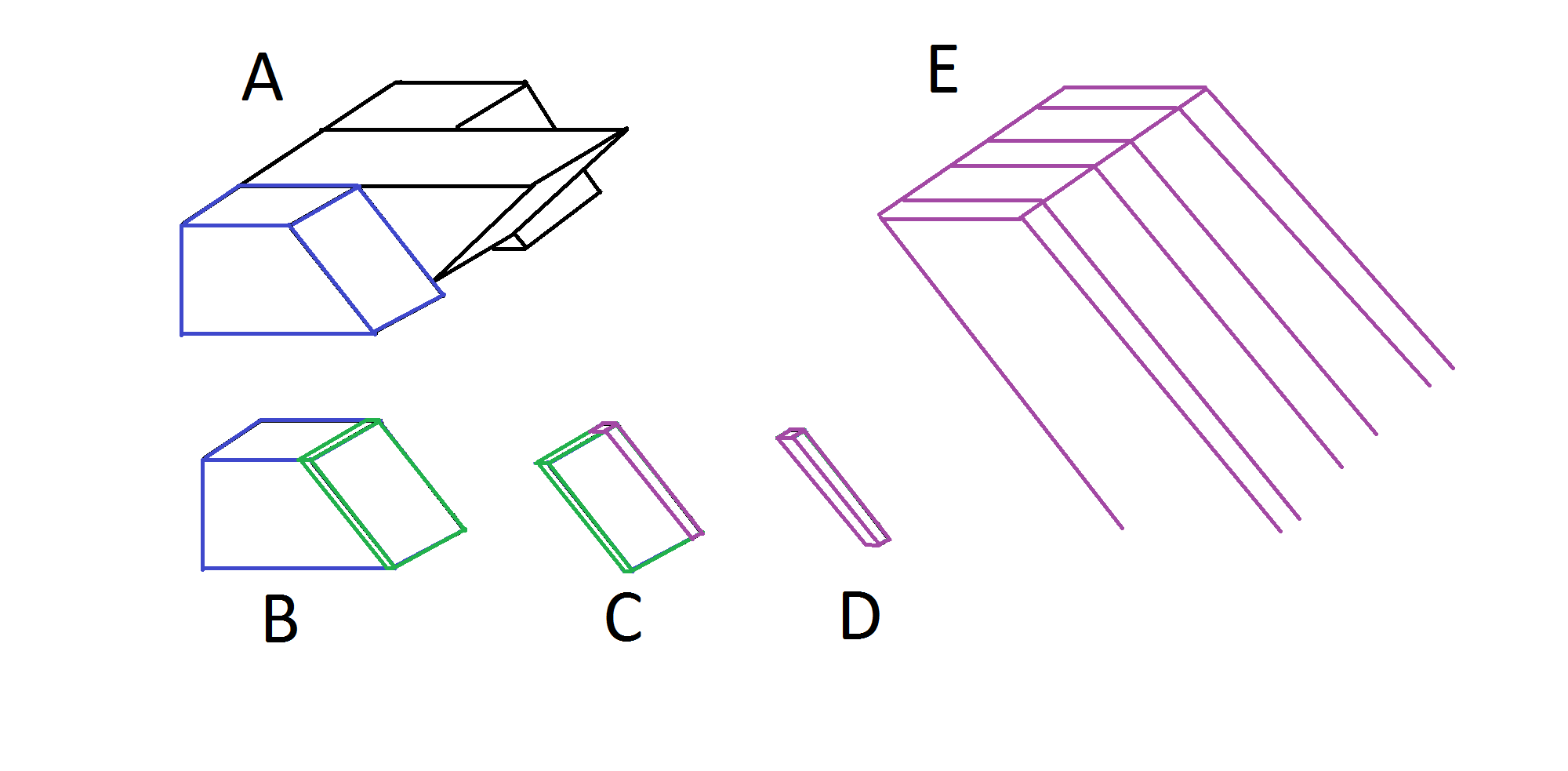 Figure 26: Diagram showing the breakdown of crossed-lamellar microstructure of molluscan shells. A: Arrangement of lamellae. B: First-order lamella. C: Second-order lamella. D: Third-order lamella. E: Banded structure of third-order lamella. Image adapted from Kobayashi and Akai (1994) [20] and Nouet et. al. (2012b) [19] by Rebecca Loh. |
Locality
Global distribution
Undate nerites have a wide distribution from Southeast Asia and northern Australia to Japan, to South Africa and Kenya, to tropical parts of North America.Figure 27: Distribution map of Nerita undata based on occurrence records available through the GBIF network, and may not represent the entire distribution. Image from EOL [5], used according to website policy. (Accessed 6 Nov 2015) |
Local distribution
Undate nerites can be found along most shores of Singapore as seen below:Habitat
Undate nerites can be found on intertidal zones (rocky shores and breakwaters) [4], and on sublittoral areas up to 3m deep [21]. They are easily found on sea walls, in crevices and under rocks [4]. Undate nerites are nocturnal, and usually emerge at dusk [14].Rearing and breeding
Undate nerites are easily bred in laboratory conditions. Adult undate nerites are easily maintained in a sheltered aquaria with constant, fresh supply of circulating seawater (personal experience). The aquaria should preferably be enclosed to prevent escapees, but not air-tight. undate nerites can be fed by providing them with algae-coated rocks, or by smearing algae (collected from seawalls or rocks) on the surface of the tank (personal experience). Undate nerites can even be self-sustainable if enough algae starts to grow on tank walls. This was confirmed by personal experience of rearing 10-20 individuals for up to three months (before they were released), during which eggs were laid.
To date, there have been no records of how to rear undate nerites in the larvae or juvenile stage, although the aquaria conditions required are probably very similar to that of adults (personal experience). The issue instead lies with how to contain these minute larvae within the tank and how to retrieve them easily.
Significance
Economic importance
Escargots? Not really. Although undate nerites are not escargots, they are known to be collected for consumption and was listed as one of the species of interest to fisheries of the Western Central Pacific [17]. However, undate nerites are not a crucial source of food for humans.The shells of undate nerites are sold online, but collecting snail shells is not recommended because of their ecological significance!
Figure 28: Screen shot of undate nerite shells on sale, here. Image by Rebecca Loh. |
Ecological significance
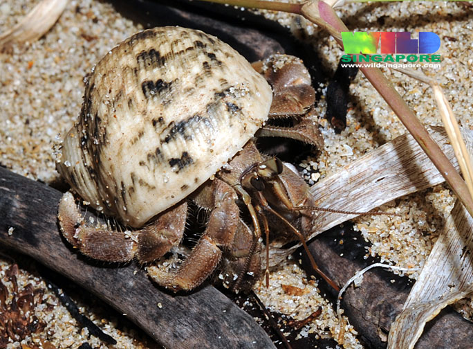
Hermit CrabsWhat happens to snail shells after they die? Before the shells degrade and minerals are returned to the environment, snail shells may act as "little houses" for a group of critters - the hermit crabs. Hermit crabs are crustaceans with fragile abdomens [22]. They inhabit empty gastropod shells and utilise them for protection. Hermit crabs will discard old shells and move into larger snail shells as they grow. A study [22] on the only species of hermit crabs found at the Red Sea - Coenobita scavola (Forskäl, 1775) - showed that 87% of the hermit crabs utilized the shells of Nerita undata. However, it was found that shell preference is not affected by the gastropod species, but instead by other factors like the shell dimensions, weight and volume etc.
Although snail shells are often sold online, collecting shells and supporting the shell trade is not recommended. Think about it - you might be depriving a hermit crab of a home!
 |
The plight of hermit crabs have inspired the name of a group of voluntary nature guides in Singapore: The Naked Hermit Crabs. The volunteers provide guided tours along the shores of Singapore, for the purpose of raising public awareness and conservation. Check out their website if you are interested in a first-hand experience with Singapore's marine biodiversity!
Environmental indicators
Although undate nerites have not been studied specifically, closely related species like Nerita lineata (Lined nerites) has been used as environmental indicators. In a study [3], the heavy metal concentrations of the shell, operculum and soft tissue of lined nerites have been used for comparison between parts of Indonesia and Malaysia successfully.
Conservation status
Fortunately, the species is neither listed in the IUCN Red List of Threatened Species, nor The Singapore Red Data Book which lists the threatened animals and plants of Singapore.
Taxonomy and systematics
Systematics
Currently, undate nerites are classified [1, 6] as below:| Kingdom |
Animalia |
||||||||
| Phylum |
Mollusca |
||||||||
| Class |
Gastropoda |
||||||||
| Subclass |
Neritimorpha |
||||||||
| Order |
Cycloneritimorpha |
||||||||
| Family |
Neritidae |
||||||||
| Genus |
Nerita |
||||||||
| Subgenus |
Cymostyla |
||||||||
| Species |
Nerita undata |
Type
A neotype for Nerita undata Linné, 1758 was designated recently, in 2006 [7]. This was to clarify the long historical confusion over its taxonomic status, which has resulted in multiple morphological variants of Nerita undata. The decision was made after finding that the original description of this species was ambiguous, the type specimen was lost and type figures were in fact figures of another well established species (with its own type). The name "Nerita undata" also could not be dropped since it was often used.The neotype was collected from the Moluccas, Indonesia (following the holotype), and is currently deposited in the Nationaal Natuurhistorisch Museum - Naturalis, Leiden, The Netherlands. The researchers have met all qualifying conditions for designating the neotype mentioned in the Code, article 75.3.
Phylogeny
The latest phylogenetic study of Genus Nerita [6] - which comprehensively sampled multiple morphological variants of Nerita undata across Asia, Southeast Asia and Australia - revealed that Nerita undata comprises multiple "evolutionarily significant units" between and within morphological variants. Distinct lineages within each morphological variant have been labeled A-C in Figure 30. Further studies are necessary to determine if these units reflect strong population genetic structure or distinct species.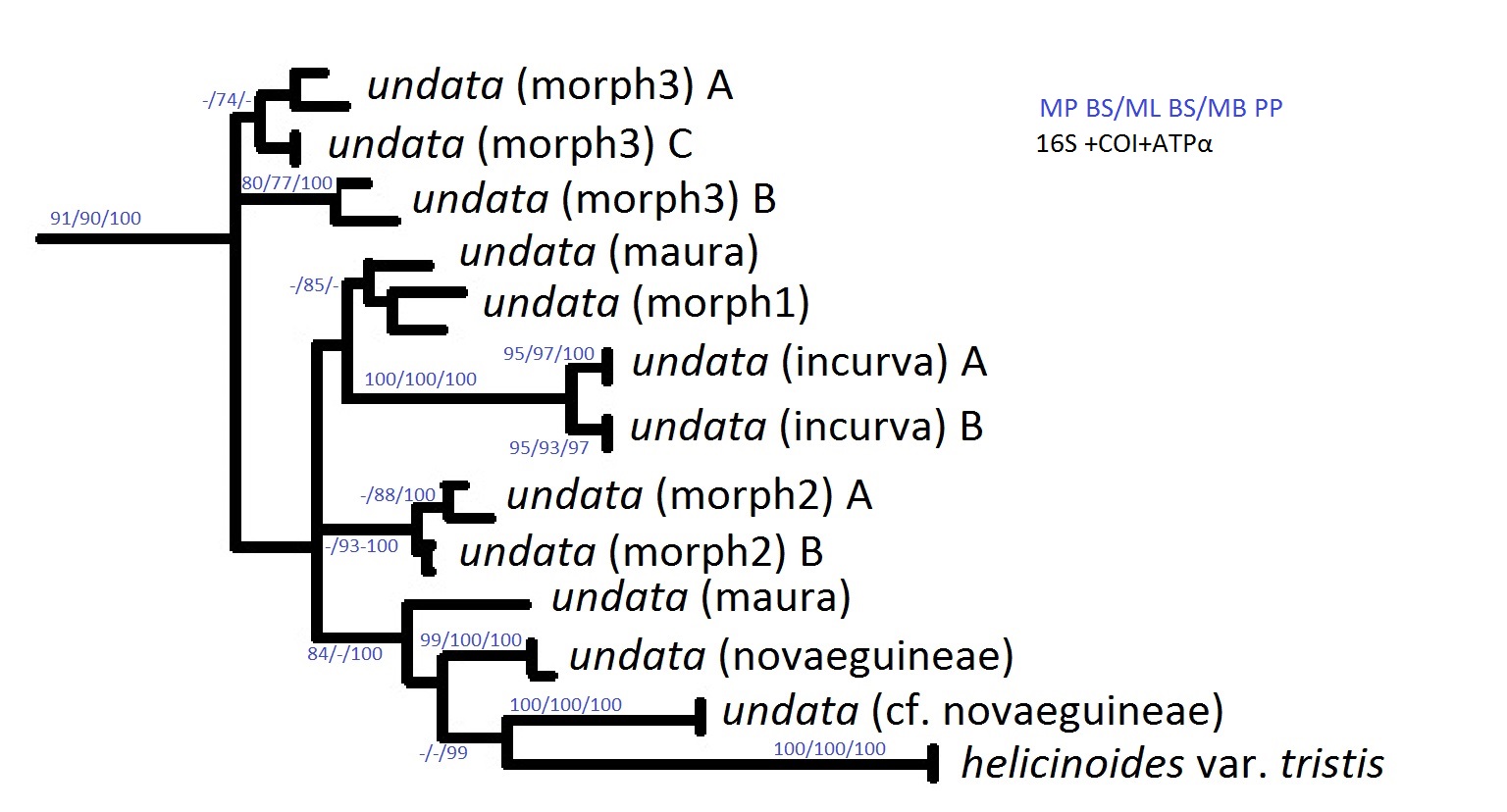 Figure 30: Molecular phylogeny of Nerita undata extracted and adapted from phylogeny of Genus Nerita. Phylogeny reconstructed from combined Bayesian analysis of mitochondrial genes (16S and COI) and nuclear genes (ATPSα). Support for each clade estimated with Parsimony (MP) and Maximum Likelihood (ML), bootstrap proportions (BS≥70%), as well as Bayesian (MB) posterior probabilities (PP≥95%). "A", "B" and "C" represent an evolutionarily significant lineage within a morphological variant. Image adapted from Frey (2010) [6] by Rebecca Loh. |
Nerita undata was also recategorised under subgenus Cymostyla von Martens, 1887, and designated as type species for Cymostyla.
Taxonomic status of local populations
A taxonomic study [4] done on the local (Singapore) populations of nerites found that undate nerites "do not show clear intraspecific variation and probably consist of a single species". Furthermore, the study confirmed that the populations in Singapore agrees with the description of the neotype designated.References
[1] "Nerita undata, Undate Nerite - names", Encyclopedia of Life. Accessed 25 Nov 2015, available from http://eol.org/pages/4881982/names/common_names
[2] "Nerita undata, Undate Nerite - details", Encyclopedia of Life. Accessed 6 Nov 2015, available from http://eol.org/pages/4881982/details
[3] "Nerita undata, Undate Nerite - details", Encyclopedia of Life, Accessed 6 Nov 2015, available from http://eol.org/pages/4881982/details
[4] Tan, S. K. & R., Clements (2008). Taxonomy and Distribution of the Neritidae (Mollusca: Gastropoda) in Singapore. Zoological Studies, 47(4):481-494
[5] "Nerita undata, Undate Nerite: Maps". In Global Diversity Information Facility. Accessed 6 Nov 2015, available from Encyclopedia of Life, http://eol.org/pages/4881982/maps
[6] Frey, M. A. (2010). A revised classification of the Gastropod Genus Nerita. The Veliger, 51(1):1-7
[7] Krijnen, C., A., Delsaerdt, N., Severijns, M., Verhaeghe & R., Vink (2006). The problematic identity of Nerita undata Linné, 1758, with designation of a neotype. (Gastropoda: Neritidae). Gloria Maris, 45(3-4):66-90
[8] Andrews, E. A. (1933). The Storage Sac For Capsule Reinforcement in Neritidae. Science, 78:39-41
[9] Houston, R. S., (1990). Reproductive Systems of Neritimorph Archaeogastropods from the Eastern Pacific, with Special Reference to Nerita funiculata Menke, 1851. The Veliger, 33: 103-110
[10] Andrews, E. A. (1937). Certain reproductive organs of Neritidae. Journal of Morphology, 61(3):525-561.
[11] Tan, K. S. & S. S., Lee (2009). Neritid egg capsules: are they all that different? Steenstrupia, 30:115-125
[12] Andrews, E. A. (1935). The egg capsules of certain Neritidae. Journal of Morphology, 57:31-59
[13] Fretter V. (1965). Functional studies of the anatomy of some neritid prosobranchs. J. Zool., 147:46-74
[14] Ruwa R.K. & V. Jaccarini (1988). Noctural feeding migrations of Nerita plicata, N. undata and N. textilis (Prosobranchia: Neritacea) on the rocky shores at Mkomani, Mombasa, Kenya. Marine Biology, 99(2):229-234
[15] Bourne, G. C. (1908). Contributions to the morphology of the group Neritacea of Aspidobranch Gastropods. Part I. The Neritidae, Proceedings of the Zoological Society of London, 1908:810-887
[16] Qi, Z. Y. (2004). Seashells of China. China Ocean Press, Beijing, pp. 418
[17] Poutiers, J.M. (1998). Gastropods. In Carpenter, K.E. and Niem, V.H. (Eds.), FAO species identification guide for fishery purposes. The living marine resources of the Western Central Pacific - Volume 1: Seaweeds, corals, bivalves and gastropods. pp. 420-430. Rome, FAO.
[18] Nouet, J., M., Cotte, J.-P., Cuif, Y., Dauphin and M., Salomé (2012a). Biochemical change at the setting-up of the crossed-lamellar layer in Nerita undata shell (Mollusca: Gastropoda), Minerals, 2(2):85-99
[19] Nouet, J., A., Baronnet, and L., Howard (2012b). Crystallization in organo-mineral micro-domains in the crossed-lamellar layer of Nerita undata (Gastropoda, Neritosina), Micron, 43:456-462
[20] Kobayashi, I., and J., Akai (1994). Twinned aragonite crystals found in the bivalvian crossed lamellar shell structure, Jour. Geol. Soc. Japan, 100(2):177-180
[21] "Nerita undata: Habitat". In Ocean Biogeographic Information System. Accessed 6 Nov 2015, available from Encyclopedia of Life, http://eol.org/data_objects/32952397
[22] Sallam, W.S, F.L., Mantelatto, and M.H., Hanafy (2008). Shell utilisation by the land hermit crab Coenobita scaevola (Anomura, Coenobitidae) from Wadi El-Gemal, Red Sea, Belg. J. Zool, 138(1):13-19
[23] Amin, B., A., Ismail, A., Arshad, C.K., Yap and M. S., Kamarudin (2006). A comparative study of heavy metal concentrations in Nerita lineata from the intertidal zone between Dumai Indonesia and Johor Malaysia, Journal of Coastal Development 10:19-32
Acknowledgements
This species page would not have been possible if not for the following people:
Mr. Tan Siong Kiat, who provided me with advice, suggestions and literature that I could not access online.
Dr. Tan Koh Siang and TMSI, for offering their facilities for me to to rear and observe the undate nerites.
Mr. Tan Zong Rui, for help with feeding my nerites and releasing them.
